Research design
The study employed an exploratory research design, with limited sites to be investigated for a minimal number of sampled bats. The aim of the study is to assess the occurrence of bat pathogens in the Malagos watershed area and characterize them in order to help identify research gaps/priorities for future studies since there is no prior information available.
Site Selection
Selection of sites for bat collection within Malagos, Davao City were done. Bats are known to frequent the area, and human or livestock exposure to bats is high because the watershed reservation is situated near residential, commercial, agricultural, industrial and tourist establishments such as the Philippine Eagle Center and Malagos Garden Resort. Malagos is also part of the Talomo Lipadas Watershed, wherein a larger research initiative on biodiversity is being explored by UP Mindanao.
Fieldwork were conducted in sampling sites established in the following conditions: a forest near human settlements; an agricultural area; and within human communities. Two sampling sites per condition will be used in the study wherein a target of 5-10 individuals will be collected per site.
GIS Mapping and Processing of Necessary Permits
Prior to sampling, permit for collection of wildlife (Gratuitous Permit) from the Regional Office of the Department of Environment and Natural Resources was secured. The study will also be subjected to evaluation by the Institutional Animal Care and Use Committee of the University of the Philippines Manila, in order to assure that the methods are in accordance with the Guidelines for the Care and Use of Laboratory Animals. GIS mapping of the sampling sites, location of the nets and possible roosting sites of bats were done. The GIS maps will be utilized for mapping the bats and their pathogens in the watershed area.
Bat Collection, Processing and Collection of Tissue and Fecal Samples
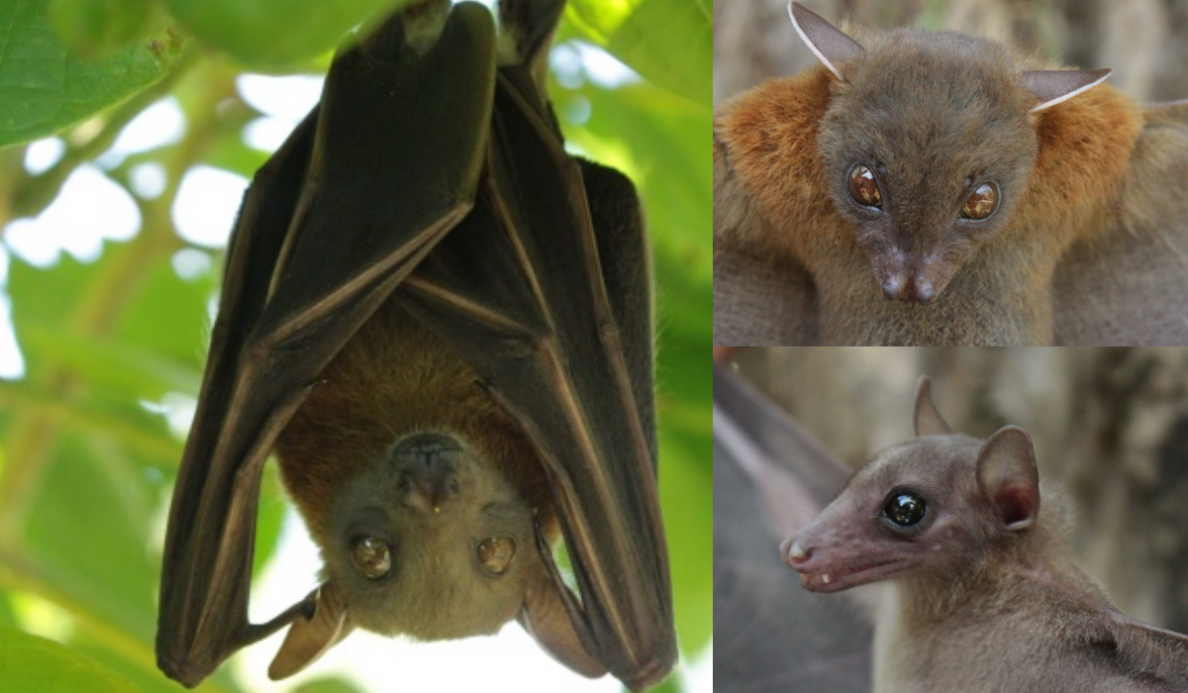
Bats were collected using convenience sampling through the mist-netting technique. Only non-endemic and non-threatened common species such as Cynopterus brachyotis, Rousettus amplexicaudatus, Macroglossus minimus and Eonycteris spelaea were included in the sampling. Mist nets were set along strategic flight paths and beside a fruit-bearing plant (for frugivorous species). These were checked regularly at three hour-intervals, and the captured animals were placed immediately in a specimen bag and processed immediately.
All captured bats were characterized on-site. The necessary biometrics were taken using caliper and ruler and was recorded in a data sheet. Other notable characteristics such as the color of fur, and the structure of the nose and interfemoral membrane were recorded. Identification of bat samples were done using the identification key of Ingle and Heaney (1992).
Euthanasia
A total of 50 individuals of non-endemic and non-threatened common bats samples were sacrificed. This was done by anesthetizing the animal through an intraperitoneal injection of tiletamine-zolazepam and killed by cardiac exsanguination (Watanabe et al, 2010).
Tissue, Urine and Fecal Collection
Blood collection was done using the cardiac puncture method while urine was aseptically collected by bladder puncture. Serum was separated and was used for MAT (Villanueva, 2010 and 2014). Kidneys were aseptically harvested and were used for Leptospira culture. Urinary bladder was aseptically removed and used for isolation of Leptospira if urine was not available. Fresh fecal samples either from the anus or within the rectum of the bat were stored in a vial and placed in an ice box. Samples were immediately transported to UP Mindanao for storage and processing.
Coronavirus Detection
RNA extraction was performed using the SV Total RNA Isolation kit (Promega, USA) using 100 mg bat small and large intestine tissue samples according to the manufacturer’s instructions. The RNA extracts were used for reverse transcription PCR (RT-PCR) using the One-step RT-PCR kit (Qiagen, USA) and published primers of Hu et al., 2018 for the amplification of the RNA-dependent RNA polymerase gene, which has been designed for the universal detection of coronaviruses. In-house primers were designed for the subsequent nested PCR reaction specific for the amplification of a 435-bp region of the RdRp gene for bat coronaviruses.
Serotyping, Isolation and Detection of Pathogenic Leptospira
Microscopic agglutination test (MAT) were performed to determine the presumptive serovar that is present in bats. Serially diluted serum samples were mixed with a panel of known Leptospira serovars and an endpoint titer giving >50% agglutination will be considered as positive (Villanueva et al., 2010).
Blood, urine or urinary bladder and kidneys were cultured in modified Korthof’s medium containing 5-fluoroucacil for the detection of pathogenic Leptospira. They were incubated at 30°C and examined weekly for leptospiral growth (Villanueva, 2010 and 2014) . Leptospires were harvested when culture is confluent and genomic DNA were collected using a commercial DNA isolation kit. Genotyping and sequencing of the bats isolate were performed by targeting the B subunit gene (gyrB) of the leptospire (Villanueva et al., 2010).
Whole kidney samples were collected and washed with saline phosphate buffer solution. The samples were subjected to genomic DNA extraction using the Blood and Tissue Kit (Qiagen, USA). Flagellin B gene (flaB) was targeted for amplification using the published primers of Kawabata et al., 2001 in order to detect and characterize Leptospira species.
Phylogenetic Analysis
PCR or RT-PCR products were extracted and sent to an outsource facility for sequencing. Reference sequences for both Leptospira and Coronaviruses were obtained from the NCBI database. Phylogenetic analysis was performed using the BEAST v.1.10.4.
Secondary data of human infections
Data on human cases of leptospirosis or coronavirus infection in Davao City were gathered through the City Health Office and Department of Health (e.g. http://www.chd11.doh.gov.ph/resu/index.php?option=com_content&task=view&id=165&Itemid=179) from year 2007 to present (10-year period). The information was used to assess the public threat of such infections in the local scene and to evaluate the relevance of the data gathered from the phylogenetic analysis of the bat pathogens.
