Emerging diseases bring attention in the public health security of the country which makes it an important focus of study in the Philippines. Increasing globalization and mobility also expands the opportunity for international spread of diseases. Consequently, the country is constantly battling against locally circulating infectious agents that recur in the population. Emerging diseases endanger not only the health of the Filipino people, but potentially damage the country’s economy as well. According to One Health Initiative, around 70% of emerging and re-emerging diseases are vector-borne or zoonotic.
Pathogenic leptospires and coronaviruses are among the emerging pathogens that threaten the Philippines. Due to its waterborne mode of transmission, leptospirosis imposes a recurring threat as cases have been reported to surge during the rainy or typhoon season (Mangunay, 2016). Though common cases of leptospirosis are usually expected after heavy rain and floods, even during the dry season, reports of leptospirosis outbreaks, particularly in agricultural areas were observed (Yanagihara et al, 2007). Davao Region was known to have the highest Leptospirosis cases reported to be in Mindanao in the year 2011 (Biliran, 2011). Typhoon Washi that hit Northern Mindanao caused an outbreak of the disease in 2012, (IFRC, 2012). Among other diseases such as Severe Acute Respiratory Syndrome (SARS) which became a global epidemic in 2003 and the Middle East Respiratory Syndrome Coronavirus (MERS-CoV) in 2012 (WHO, 2003 and 2019), lethal respiratory diseases caused by members of the Coronavirus family has not spared the Philippines to be drastically affected.
Bats act as natural reservoirs of infectious agents and has gained tremendous scientific interest in the field of disease ecology. Experts warned that a number of zoonotic and potentially zoonotic pathogens are known to circulate in bats, and anthropogenic activities that increase exposure to bats will favor ecological spillover of such pathogens to the environment and humans (Hayman et al., 2013). Wild land disturbance and demographic changes, according to Peter Daszak, were known to be the causes of diseases that have emerged from the last 30 or 40 years (Robbins, 2012). Hence, public health experts are starting to recognize the role of ecology in emerging diseases. One Health Initiative would be a great example, a worldwide program that acknowledges the inextricable relationship between human, animal and ecological health, and advocates for a holistic approach in health research (One Health Initiative, n.d.).
Significance of the study
The occurrence of leptospires and coronaviruses in bats, their phylogenetic relationship with human strains, and their distribution in terms of locations were the main focus of this study. Highly diverse strains of Leptospira have been detected in bats, especially in areas with high bat species richness (Dietrich, 2014; Lagadec, 2012; and Matthias, 2005). However, as stated earlier, it still remains uncertain if bats serve as sources of pathogenic strains transmitted to humans and susceptible animals.
There is increasing evidence for the bat origin of SARS-CoV and MERS-CoV. Both have demonstrated close genetic relationship with coronaviruses isolated from bats (Hu et al, 2015). Only a couple of studies have investigated locally circulating bat coronaviruses in the Philippines (Watanabe et al., 2010), and, to date, none for bat leptospires. This highlights a huge opportunity for health research.
Bats samples were collected from Malagos, Davao City, a barangay situated near a watershed reservation along with residential, commercial, agricultural, and industrial establishments. It is also home to the Philippine Eagle Center and Malagos Garden Resort. This area is known to be frequented by bats. An information system will be created to help organize and manage the data, and can be made accessible to the public in the future. This information will be useful for public health stakeholders to help identify the strains and hotspot sources of local outbreaks caused by ecological spillovers.
Bats and their role as natural reservoir of pathogens
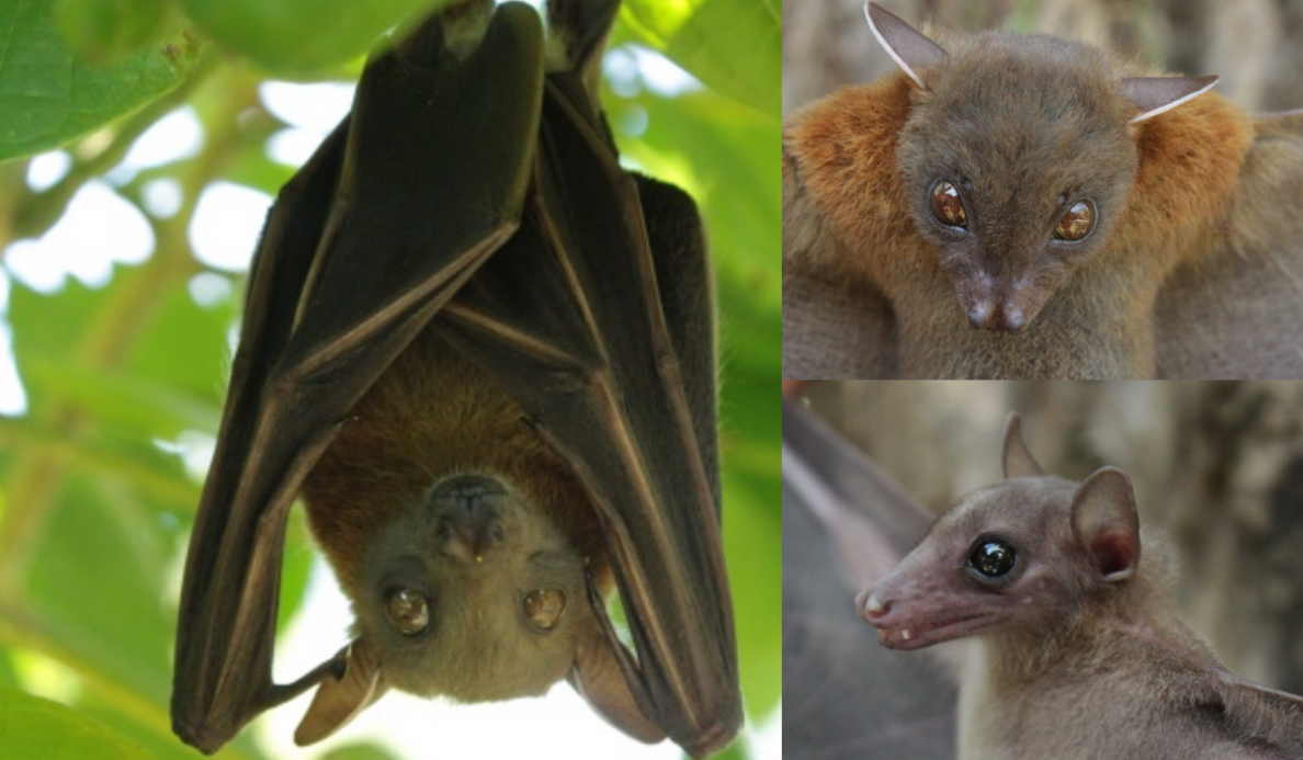
With at least 73 known species, bats are one of the most diverse group of mammals in the Philippines (Heaney and Regalado, 1998; Heaney et al., 1998). There are over 53 species which includes fruit bats and insect bats in Mindanao (Ingle et al., 1999). They occur from primary forests to disturbed forests in the country (Heaney et al., 1998). Agricultural lands, villages and cities were among the ecosystems these several species are known to co-inhabit. Fruit bats such as Cynopterus brachyotis, Macroglossus minimus and Rousettus amplexicaudatus were among the fruit bats known to occur in areas within human communities (Ingle et al., 1992). Insect bats such as Scotophilus kuhlii were also commonly recorded roosting in buildings and other urban infrastructures (Heaney et al., 1998).
These flying animals may serve important roles in the ecosystem and the economy. Bats are good agents of seed dispersal, pollinate high valued crops and limits the population of some insect pests (Kasso and Balakrishnan, 2013). However, emerging viruses such as rabies and Nipah virus may also serve these bats are reservoir hosts (Calisher et al., 2006).
Laboratory Diagnostics
Several diagnostic methods for leptospirosis that are being routinely used include:
Direct examination by dark field microscopy
Dark Field microscopy is a microscope illumination technique used to observe unstained samples causing them to appear brightly lit against a dark, almost purely black, background. Rays are scattered in all directions when light hits an object. The design of the dark field microscope is such that it removes the dispersed light so that only the scattered beams hit the sample. This type of microscopy is ideal for viewing objects that are unstained, transparent and absorb little or no light. In studying marine organisms such as algae and plankton, diatoms, insects, fibres, hairs, yeast, live bacterium, protozoa as well as cells and tissues, dark field microscopy is also used. It is ideal for live blood analysis enabling the practitioner to see much more than is possible with other lighting methods (Clemons, 2016).
Polymerase Chain reaction (PCR)
PCR is based on using the ability of DNA polymerase to synthesize new strand of DNA complementary to the offered template strand. It needs a primer to which it can add the first nucleotide because DNA polymerase can add a nucleotide only onto a preexisting 3′-OH group. This requirement makes it possible to delineate a specific region of template sequence that the researcher wants to amplify. The specific sequence will be accumulated in billions of copies (amplicons) at the end of the PCR reaction. (National Center for Biotechnology Information, 2017)
Components of PCR
DNA template: This is the sample DNA that contains the target sequence. High temperature is applied to the original double-stranded DNA molecule to separate the strands from each other at the beginning of the reaction.
DNA polymerase: This is a type of enzyme that synthesizes new strands of DNA complementary to the target sequence. The first and most commonly used of these enzymes is TaqDNA polymerase (from Thermis aquaticus), whereas PfuDNA polymerase (from Pyrococcus furiosus) is used widely because of its higher fidelity when copying DNA. Although these enzymes are subtly different, they both have two capabilities that make them suitable for PCR namely: 1) using a DNA template and primers, they can generate new strands of DNA, and 2) they are heat resistant.
Primers: These are short pieces of single-stranded DNA that are complementary to the target sequence. The polymerase begins synthesizing new DNA from the end of the primer.
Nucleotides (dNTPs or deoxynucleotide triphosphates): These are single units of the bases A, T, G, and C, which are essentially “building blocks” for new DNA strands.
RT-PCR (Reverse Transcription PCR) is PCR preceded with conversion of sample RNA into cDNA with the enzyme reverse transcriptase (National Center for Biotechnology Information, 2017)
Microscopic Agglutination Test
Microscopic agglutination test (MAT) has been widely used as the reference test for antibody detection. MAT is performed by incubating patient serum with various serovars of leptospires. MAT titer is obtained by testing various serum dilutions with the positive serovar. The serovar that reacts with patient serum is suggested to be the infecting serovar. Information on infecting serovars obtained by MAT has been used for epidemiological study. (Chirathaworn et al. 2014)
ELISA – Enzyme-linked Immunosorbent Assay
ELISA tests are utilized to detect substances that have antigenic properties, primarily proteins. Some of these include hormones, bacterial antigens and antibodies. (Shiel, 2018)
Immunoglobulins such as IgG and IgM antibodies and chemicals for the detection of immune responses in the body are the components used in an ELISA test. The ELISA test involves an enzyme. It also involves an antibody or antigen that may form an antigen-antibody reaction to provide a positive result or, if they do not react, a negative result. Examples of the uses of an ELISA test include diagnosing infections such as HIV (human immunodeficiency virus) and some allergic diseases like food allergies and experimental investigations to identify compounds.
The test is based on a microtiter plate that has a solid phase substrate (target protein, antigen) at a known concentration fixed to the plate that when exposed to an antibody that has an indicator attached (dye for color change or enzyme-labeled antibody) that can produce a color change.
Types of ELISA
Direct ELISA: attachment of an antigen to a polystyrene plate followed by an enzyme-labeled antibody that can react with the antigen and a substrate that can be measured.
Indirect ELISA: attachment of an antigen to a polystyrene plate followed by an unlabeled or primary antibody followed by an enzyme-labeled antibody that can react with both the primary antibody and substrate
Sandwich ELISA: A capture antibody is attached to the polystyrene plate, then antigen is added that specifically attaches or captures the antigen. A second antibody, also specific for the antigen but not the same as the capture antibody is added and “sandwiches” the antigen. This second antibody is then followed by an enzyme-labeled antibody specific for the second antibody that can react with a substrate that can be measured
Competitive ELISA: This test is like the sandwich ELISA but involves the addition of competing antibodies or proteins when the second antibody is added. This results in a decrease in the substrate signal generated. This test gives good, highly specific results.
ELISA in detection of Leptospira
This test detects antibodies to LipL32, a membrane protein found on pathogenic leptospires. The currently available assay provides a qualitative negative or positive result and will also detect antibodies induced by vaccination. A comparison of this test to the MAT has not been reported, and it is likely that the numerical titers provided by the MAT will provide more diagnostically useful information than a qualitative ELISA. (Lunn, 2016)
In performing ELISA, an antigen must be immobilized on a solid surface and then complexed with an antibody that is linked to an enzyme. Detection is accomplished by assessing the conjugated enzyme activity via incubation with a substrate to produce a measurable product. The most crucial element of the detection strategy is a highly specific antibody-antigen interaction. (Thermo Fisher, n.d.)
RT-PCR
RT-PCR (Reverse Transcriptase Polymerase Chain Reaction) is commonly used to test for genetic diseases and to characterize gene expression in various tissue types, cell types, and over developmental time courses. This serves as a form of expression profiling, but typically as a candidate approach. RT-PCR is also commonly used to clone cDNAs for further use with other molecular biology techniques. (Bachman, 2013)
RT-PCR allows the use of RNA as a template. An additional step allows the detection and amplification of RNA. Using reverse transcriptase, the RNA is reverse transcribed into complementary DNA (cDNA). The quality and purity of the RNA template is essential for the success of RT-PCR. The first step of RT-PCR is the synthesis of a DNA/RNA hybrid. Reverse transcriptase also has an RNase H function, which degrades the RNA portion of the hybrid. The single stranded DNA molecule is then completed by the DNA-dependent DNA polymerase activity of the reverse transcriptase into cDNA. The efficiency of the first-strand reaction can affect the amplification process. From here on, the standard PCR procedure is used to amplify the cDNA. The possibility to revert RNA into cDNA by RT-PCR has many advantages. RNA is single-stranded and very unstable, which makes it difficult to work with. Most commonly, it serves as a first step in qPCR, which quantifies RNA transcripts in a biological sample. (Neidler, 2019)
Comparison of ELISA and RT-PCR
Lau et al. (2005) compared two methods for detecting CoVs: ELISA and reverse transcription-PCR (RT-PCR). Results are produced within hours with ELISA and it is also less expensive and easier to conduct. However, sensitivity-wise, RT-PCR assays are generally more reliable. Lau and his team reported that RT-PCR was more sensitive than ELISA and could be used for early detection of the SARS-CoV in fecal samples of patients. Various primers have been designed based on highly conserved regions of the RNA-dependent RNA polymerase (RdRp) gene to detect both alphacoronaviruses and betacoronaviruses using RT-PCR. Woo et al. (2005) and Poon et al. (2003) developed primers for human CoV detection, while Watanabe et al. (2010) and Tsuda et al. (2012) used primers for bat CoVs in the Philippines.
Immunofluorescence Assay
Classically defined as a procedure in detecting antigens in cellular contexts using antibodies, immunofluorescence is an assay which is used primarily on biological samples. The base for immunofluorescence is the specificity of antibodies to their antigen. The biological samples include tissue and cells. Immunofluorescence allows researchers to evaluate whether or not cells in a particular sample express the antigen in question. In cases where an immunopositive signal is found, immunofluorescence also allows researchers to determine which subcellular compartments are expressing the antigen. Immunofluorescence can be used on cultured cell lines, tissue sections, or individual cells. (Sino Biological, 2019)
There are two different immunofluorescence assay which include indirect immunofluorescence assay and direct immunofluorescence assay. For indirect immunofluorescence assay, the protocol mainly include tissue or cell preparation, tissue or cell fixation, serum blocking, primary antibody incubation, marked second antibody incubation, staining, result judgment and imaging. For direct immunofluorescence assay, there are only marked primary antibody that has been incubated without second antibody and other steps are the same. (Sino Biological, 2019)
Hemmaglutination Assay
Hemagglutination is used for the diagnosis of some enveloped viruses such as influenza viruses. This method relies on the specific feature of some enveloped viruses that can adsorb to red blood cells (RBCs). Hemagglutinin5 (HA),specifically, an envelope glycoprotein of some enveloped viruses, imparts this property. By gravity, RBCs precipitate to the bottom of the well, giving rise to a distinct red-colored dot in a conical shaped well in the absence of virus particles. As a result of interaction between HA proteins of virus particles and RBC, RBCs clump together leading to a lattice formation in the presence of virus particles. In this case, as RBCs are dispersed as a clump, a red dot is not formed. In a given sample, a red dot will appear beyond a certain dilution fold. To carry out a hemagglutination assay, a twofold serial dilution of virus-containing samples is dispensed into individual wells of a 96-well microtiter plate. Then, aliquots of RBC are added to each well. The highest dilution at which clumping is observed is regarded as the HA titer of the sample. The virus titer in a sample can be estimated by multiplying the dilution fold. 1 HA unit corresponds to 104 particles per mL in a standard condition. One outstanding advantage of this method is that it does not require any equipment. Moreover, it is a robust and fast diagnostic tool, but there are limitations sensitivity-wise. (Wang-Shick, 2017)
Test Sensitivities
MAT is the most widely used reference method for the diagnosis of human leptospirosis with a sensitivity of 76-91.4% and specificity of 86.7-97% during the convalescent stage of the illness (Cumberland et al., 1999 and Eugene et al., 2015). According to WHO and ILS, it is also known as the gold standard for serological diagnosis of leptospirosis (WHO and ILS, 2003). Commercial IgM ELISAs have a sensitivity and specificity of 86.5% and 97%, respectively (Bajani et al., 2003). The sensitivity of the immunofluorescence assay was reported to be 45.5% and 91.9% and the specificity was 96.8 and 100% (Kemapunmanus et al., 2004 and Limmathurotsakul, 2012). Finally, the haemagglutination assay was reported to have a sensitivity and specificity of 79% and 81.1%, respectively (Bajani et al., 2003).
Malagos Watershed
Under proclamation No. 612, Malagos watershed is one of the proclaimed watersheds in the country. It is located in Baguio District, Davao City and is part of the Panigan-Tamugan and Talomo-Lipadas Watersheds. The area is also known for its tourist destination such as the Philippine Eagle Center and Malagos Garden Resort. It is inhabited by locals with an increasing population trend from 1,494 in 1970s to 5,933 in the year 2010 (Ibañez et al., 2012).
In terms of biodiversity, the watershed is considered abundant. Several studies conducted within the watershed recorded diverse species of wildlife. There were 54 species of birds (Alviola et al., 2010), 9 species of amphibians (Lorica, 2009) and 11 rodent species (Aloy, 2001). In 2001, Oliva also recorded 5 species of bats within the area which includes the endemic and threatened Acerodon jubatus (Giant golden-crowned flying fox). Moreover, the near threatened-Pteropus vampyrus (Large Flying fox) was also recorded roosting within the forest of the watershed (Cayunda et al., 2004). The bat species, Cynopterus brachyotis, which is known to co-inhabit with humans were also recorded and was found to be one of the most abundant in the area (Oliva, 2001).
