
Source: Peiris, Joseph S Malik
https://researchgate.net
Virology
When viewed under a microscope, coronaviruses are a group of viruses that have a halo or crown-like (corona) appearance. They are about 100nm in diameter, are the largest positive strand RNA viruses (indeed they have the largest genomes of any RNA virus). They infect humans and animals in which they cause respiratory and enteric disease. The coronaviruses, belong to a group, the nidovirales, that produce a nested set of mRNA with a common 3’ end. The coronaviruses and the toroviruses (which together make up the Coronaviridae) have helical nucleocapsids while the arteriviruses have icosahedral nucleocapsids. Coronaviruses have an envelope that is derived from intracellular membranes and not the plasma membrane. (Hunt, 2016)
The genome RNA is complexed with the basic nucleocapsid (N) protein to form a helical capsid found within the viral membrane. The membranes of all coronaviruses contain at least three viral proteins. These are:
- spike (S), the type I glycoprotein that forms the peplomers on the virion surface, giving the virus its corona- or crown-like morphology in the electron microscope
- the membrane (M) protein, a protein that spans the membrane three times and has a short N-terminal ectodomain and a cytoplasmic tail; and
- small membrane protein (E), a highly hydrophobic protein
The E protein of IBV has a short ectodomain, a transmembrane domain, and a cytoplasmic tail (Corse and Machamer, 2000). The E protein of MHV is reported to span the membrane twice, such that both N and C termini are on the interior of the virion (Maeda et. al, 2001). Hemagglutinin esterase (HE) is an additional membrane protein known to be found in some group II coronaviruses have an additional membrane protein. (Brian, Houge and Kienzle, 2000). HE is not an essential protein and while its function is still unknown, it has been speculated to help in viral entry and/or pathogenesis in vivo. HE is not encoded in the SARS-CoV genome. There is an additional group II virion protein called I for internal, as it is encoded within the nucleocapsid open reading frame (ORF). This is a nonessential protein of unknown function (Fischer et al, 1997). It has recently been shown that the ORF 3a-encoded SARS protein is an additional structural protein (Ito et al, 2005). There may be other minor proteins, as yet undetected, included in virions.
Epidemiology
Coronaviruses are common throughout the world. There are six different coronaviruses that scientists know of which can infect people and make them sick. Some coronaviruses have been around a long time and commonly cause mild to moderate illness in people worldwide. MERS-CoV and SARS-CoV are the two newer coronavirus that have been known to frequently cause severe illness. (Centers for Disease Control and Prevention, 2017)
In 2002-2003 in Guangdong, China, an outbreak of SARS-CoV occurred and the identified causative agent is the group 2b β-coronavirus. Among the coronaviruses, it is the most severe disease. During the 2002–2003 outbreak approximately 8098 cases occurred with 774 deaths, resulting in a mortality rate of 9%. This rate was much higher in elderly individuals, with mortality rates approaching 50% in individuals over 60 years of age. The outbreak began in a hotel in Hong Kong and ultimately spread to more than two dozen countries. Closely related viruses were isolated from several exotic animals including Himalayan palm civets and raccoon dogs during the epidemic (Guan et al 2003). However, it is widely accepted that SARS-CoV originated in bats as a large number of Chinese horseshoe bats contain sequences of SARS-related CoVs and contain serologic evidence for a prior infection with a related CoV (Lau et al and Li et al, 2005). To date, two novel bat SARS-related CoVs were recently identified that are more similar to SARS-CoV than any other virus identified (Ge, 2013). Providing further evidence that SARS-CoV originated in bats. They were also found to use the same receptor as the human virus, angiotensin converting enzyme 2 (ACE2). Although some human individuals within wet animal markets had serologic evidence of SARS-CoV infection prior to the outbreak, apparently these individuals were asymptomatic (Guan, 2003). Thus, before a series of factors facilitated its spread into the larger population, it is likely that a closely related virus circulated in the wet animal markets for several years.
In 2012, Jordan was found to have the first known cases of Middle East respiratory syndrome (MERS), associated with infection by a novel coronavirus (CoV), but were reported retrospectively and it was from Jeddah, Kingdom of Saudi Arabia the case was first to be publicly reported. MERS-CoV sequences have been found in a bat and in many dromedary camels (DC) since then. MERS-CoV is enzootic in DC across the Arabian Peninsula and in parts of Africa, causing mild upper respiratory tract illness in its camel reservoir and sporadic, but relatively rare human infections. It still remains unknown how the virus can be transmitted to humans but close and lengthy exposure appears to be a requirement. The KSA is the focal point of MERS, with the majority of human cases. MERS is mostly known as a lower respiratory tract (LRT) disease involving fever, cough, breathing difficulties and pneumonia that may progress to acute respiratory distress syndrome, multiorgan failure and death in 20 % to 40 % of those infected in humans. However, MERS-CoV has also been detected in mild and influenza-like illnesses and in those with no signs or symptoms. Older males most obviously suffer severe disease and MERS patients often have comorbidities. Another sometimes- fatal zoonotic coronavirus disease that has since disappeared, MERS progresses more rapidly to respiratory failure and acute kidney injury (it also has an affinity for growth in kidney cells under laboratory conditions), is more frequently reported in patients with underlying disease and is more often fatal compared to severe acute respiratory syndrome (SARS). With approximately 20 % of all virus detections reported among healthcare workers (HCWs) and higher exposures in those with occupations that bring them into close contact with camels, most human cases of MERS have been linked to lapses in infection prevention and control (IPC) in healthcare settings. Sero-surveys have found widespread evidence of past infection in adult camels and limited past exposure among humans. Almost from the start of the emergence of MERS, sensitive, validated reverse transcriptase real-time polymerase chain reaction (RT-rtPCR)-based diagnostics have been available. While the basic virology of MERS-CoV has advanced over the past three years, understanding of the interplay between camel, environment, and human remains limited. (Mackay and Arden, 2015)
Pathogenesis
Animal Coronaviruses
In the last half of the 20th century, coronaviruses cause a large variety of diseases in animals, and their ability to cause severe disease in livestock and companion animals such as pigs, cows, chickens, dogs and cats led to significant research on these viruses. The most common are Transmissible Gastroenteritis Virus (TGEV) and Porcine Epidemic Diarrhea Virus (PEDV) cause severe gastroenteritis in young piglets, leading to significant morbidity, mortality, and ultimately economic losses. For the first time, PEDV recently emerged in North America causing significant losses of young piglets. Porcine hemagglutinating encephalomyelitis virus (PHEV) mostly leads to enteric infection but has the ability to infect the nervous system, causing encephalitis, vomiting and wasting in pigs. Feline enteric coronavirus (FCoV) causes a mild or asymptomatic infection in domestic cats, but during persistent infection, mutation transforms the virus into a highly virulent strain of FCoV (Feline Infectious Peritonitis Virus, FIPV), that leads to development of a lethal disease called feline infectious peritonitis (FIP). With similarities to the human disease, sarcoidosis, FIP has wet and dry forms. FIPV is macrophage tropic and it is believed that it causes aberrant cytokine and/or chemokine expression and lymphocyte depletion, resulting in lethal diseases (Perlman and Netland, 2009). However, additional research is needed to confirm this hypothesis. Bovine CoV, Rat CoV, and Infectious Bronchitis Virus (IBV) cause mild to severe respiratory tract infections in cattle, rats, and chickens, respectively. Bovine CoV causes significant losses in the cattle industry and also has spread to infect a variety of ruminants, including elk, deer and camels. The virus causes diarrhoea (‘winter dysentery’ and ‘shipping fever’), all leading to weight loss, dehydration, decreased milk production, and depression in addition to severe respiratory disease. (Perlman and Netland, 2009). Some strains of IBV, a γ-coronavirus, also affect the uro-genital tract of chickens causing renal disease. IBV significantly diminishes egg production and weight gain, causing substantial losses in the chicken industry each year (Perlman and Netland, 2009). A novel coronavirus named SW1 was identified in a deceased Beluga whale (Mihindukulasuriya et al, 2008). Large numbers of virus particles were identified in the liver of the deceased whale with respiratory disease and acute liver failure. Sequencing of the liver tissue clearly identified the virus as a coronavirus although the electron microscopic images were not sufficient to identify it as coronavirus. Based on the phylogenetic analysis, it was subsequently determined to be a γ-coronavirus but it has not yet been verified experimentally that this virus is actually a causative agent of disease in whales. There has also been an intense interest in identifying novel bat CoVs, since these are the likely ultimate source for SARS-CoV and MERS-CoV, and hundreds of novel bat coronaviruses have been identified over the past decade (He et al, 2014). Lastly, Mesoniviridae, another novel group of nidoviruses, were recently identified as the first nidoviruses to exclusively infect insect hosts (Nga et al, 2011 and Lauber et al, 2012). These viruses are highly divergent from other nidoviruses but are most closely related to the roniviruses. They are ∼20 kb in size, falling in between large and small nidoviruses. Interestingly, these viruses do not encode for an endoribonuclease, which is present in all other nidoviruses. These attributes suggest these viruses are the prototype of a new nidovirus family and may be a missing link in the transition from small to large nidoviruses.
The most heavily studied animal coronavirus is murine hepatitis virus (MHV), which causes a variety of outcomes in mice, including respiratory, enteric, hepatic, and neurologic infections. These infections often serve as highly useful models of disease. For instance, MHV-1 causes severe respiratory disease in susceptible A/J and C3H/HeJ mice, A59 and MHV-3 induce severe hepatitis, while JHMV causes severe encephalitis. Levy et al (2000) explained that MHV-3 induces cellular injury through the activation of the coagulation cascade. Most notably, A59 and attenuated versions of JHMV cause a chronic demyelinating disease that bears similarities to multiple sclerosis (MS), making MHV infection one of the best models for this debilitating human disease. Lampert (1973) and Weiner (1973) presented that early studies suggested that demyelination was dependent on viral replication in oligodendrocytes in the brain and spinal cord; however, more recent reports clearly demonstrate that the disease is immune-mediated. Irradiated mice or immunodeficient (lacking T and B cells) mice do not develop demyelination, but addition of virus-specific T cells restores the development of demyelination (Wu et al 1990 and Wang et al, 2000). According to Wu and Perlman, demyelination is also accompanied by a large influx of macrophages and microglia that can phagocytose infected myelin although it is unknown what the signals are that direct immune cells to destroy myelin. Lastly, MHV can be studied under BSL2 laboratory conditions, unlike SARS-CoV or MERS-CoV, which require a BSL3 laboratory, and provides a large number of suitable animal models. These factors make MHV an ideal model for studying the basics of viral replication in tissue culture cells as well as for studying the pathogenesis and immune response to coronaviruses.
Human Coronaviruses
Coronaviruses were only thought to cause mild, self-limiting respiratory infections in human prior to SARS-CoV outbreak. Two of these human coronaviruses are α-coronaviruses (HCoV-229E and HCoV-NL63) while the other two are β-coronaviruses (HCoV-OC43 and HCoV-HKU1). McIntosh Bradburne and Hamre et al in their studies, HCoV-229E and HCoV-OC43 were isolated nearly 50 years ago while HCoV-NL63 and HCoV-HKU1 were only recently identified following the SARS-CoV outbreak (Woo et al, 2005 and van der Hoek, 2004). Causing 15-30% of respiratory tract infections each year, these viruses are endemic in human populations. They cause more severe disease in neonates, the elderly, and in individuals with underlying illnesses, with a greater incidence of lower respiratory tract infection in these populations. One interesting aspect of these viruses is their differences in tolerance to genetic variability. According to Chibo and Birch (2006), HCoV-229E isolates from around the world have only minimal sequence divergence while HCoV-OC43 isolates from the same location but isolated in different years show significant genetic variability according to Vijgen et al (2005). This likely explains the inability of HCoV-229E to cross the species barrier to infect mice while HCoV-OC43 and the closely related bovine coronavirus, BCoV, are capable of infecting mice and several ruminant species. It has been suggested that human CoVs may be involved in the development of multiple sclerosis (MS) based on the ability of MHV to cause demyelinating disease. However, no evidence to date suggests that human CoVs play a significant role in MS.
Epithelial cells within the lung are the primary site of infection of the SARS-CoV. The virus is capable of entering macrophages and dendritic cells but only leads to an abortive infection (Peiris et al, 2003 and Spiegel et al, 2006). The infection of these cell types may also be important in inducing pro-inflammatory cytokines that may contribute to disease (Law et al, 2005). Numerous cytokines and chemokines in fact are produced by these cell types and are elevated in the serum of SARS-CoV infected patients (Lau and Peiris, 2005). It still remains unknown the exact mechanism of lung injury and cause of severe disease in humans remains. When severe disease develops in both humans and in several animal models of the disease, viral titers seem to diminish. Furthermore, animals infected with rodent-adapted SARS-CoV strains show similar clinical features to the human disease, including an age-dependent increase in disease severity (Roberts et al, 2005). The levels of proinflammatory cytokines were increased and T-cell responses were reduced in these animals which suggests a possible immunopathological mechanism of disease (Zhao et al 2010 and 2011)
Diagnosis
In diagnosing coronavirus, the specimen that can be used are respiratory secretions, and stool (HKU1). (Vabret et. al, 2006)
Virus isolation
The human hepatoma cell-line HUH7 has been recently used for primary isolation of OC43, 229E and HKU-1 viruses from clinical specimens and NL63 has been isolated in LLC-MK2 and Vero B4 cell. (Aryal, 2019)
RT-PCR
Another way to detect coronavirus viral RNA is by RT-PCR. Reverse transcriptase polymerase chain reaction is the technology by which RNA molecules are converted into their complementary DNA (cDNA) sequences by reverse transcriptases, followed by the amplification of the newly synthesized cDNA by standard PCR procedures. The role of reverse transcriptase (RT) in the synthesis of first-strand cDNA is the primary component of this technology. RT-PCR is a two-step process. Using some variant of PCR, it involves reverse transcription of purified RNA by RT via an appropriate method for priming and amplification of first-strand cDNA. Normalization of samples is very important in RT-PCR, and the efficiency of first-strand cDNA synthesis is one of the most important determinants in the success or failure of this method. Rather than repeating the same cDNA synthesis over and over, it is strategically better to make a large cDNA pool from which aliquots may be drawn for individual applications. It is necessary to have useful primers designed in promoting a proper balance between template specificity, thermodynamic stability when base-paired to the template, and capacity of one primer to function with the other(s) to support RT-PCR. The probable collaborative behavior of one or more pairs of oligonucleotide primers is best described in terms of the Tm of each primer involved. The Tm is that temperature at which 50% of the possible annealing events between primer and template have occurred and 50% have yet to occur. (Farrel, 2010)
Electron microscopy of negatively stained stool specimens is useful for the detection of enteric coronaviruses. (Aryal, 2019)
Complement fixation, ELISA assays, immunofluorescence or virus neutralization tests have been used for serological diagnosis. (Aryal, 2019)
Serologic diagnosis of infections with strain 229E is possible using a passive hemagglutination test in which red cells coated with coronavirus antigen are agglutinated by antibody-containing sera. (Aryal, 2019)
HCoV-OC43–related virions that express an HE glycoprotein on the viral envelope can also be detected by hemagglutination and acetyl esterase assays. (Aryal, 2019)
Prevention
Currently, there are no vaccines available to protect you against human coronavirus infection. You may be able to reduce your risk of infection by doing the following:
- wash your hands often with soap and water
- avoid touching your eyes, nose, or mouth with unwashed hands
- avoid close contact with people who are sick
(Centers for Disease Control and Prevention, 2017)
Transmission
The SARS coronavirus (SARS Co-V) is predominantly spread in droplets that are shed from the respiratory secretions of infected persons. Fecal or airborne transmission seem to be less frequent. (Kamps and Hoffman, 2006)
According to Centers for Disease Control and Prevention (2017), the spread of common human coronaviruses have not been studied very much. However, human coronaviruses likely spread from an infected person to others through:
- the air by coughing and sneezing
- close personal contact, such as touching or shaking hands
- touching an object or surface with germs on it, then touching your mouth, nose, or eyes before washing your hands
- rarely, fecal contamination
In the United States, people usually get infected with common human coronaviruses in the fall and winter. However, you can get infected at any time of the year. Most people will get infected with one or more of the common human coronaviruses in their lifetime. Young children are most likely to get infected. However, people can have multiple infections in their lifetime.
On the other hand, MERS-CoV, like other coronaviruses, likely spreads from an infected person’s respiratory secretions, such as through coughing. However, same with SARS, it is not fully understood the precise ways how the virus spreads.
MERS-CoV has spread from sick people to others through close contact, such as caring for or living with an infected person. Infected people have spread MERS-CoV to others in healthcare settings, such as hospitals. Researchers studying MERS have not seen any ongoing spreading of MERS-CoV in the community.
All reported cases have been linked to countries in and near the Arabian Peninsula. Before they contracted the disease, most infected people either lived in the Arabian Peninsula or recently traveled from the Arabian Peninsula. A few people became infected with MERS-CoV after having close contact with an infected person who had recently traveled from the Arabian Peninsula. The largest known outbreak of MERS outside the Arabian Peninsula occurred in the Republic of Korea in 2015. It was associated with a traveler returning from the Arabian Peninsula.
Public health agencies continue to investigate clusters of cases in several countries to better understand how MERS-CoV spreads from person to person. (Centers for Disease Control and Prevention, 2017)
Zoonosis
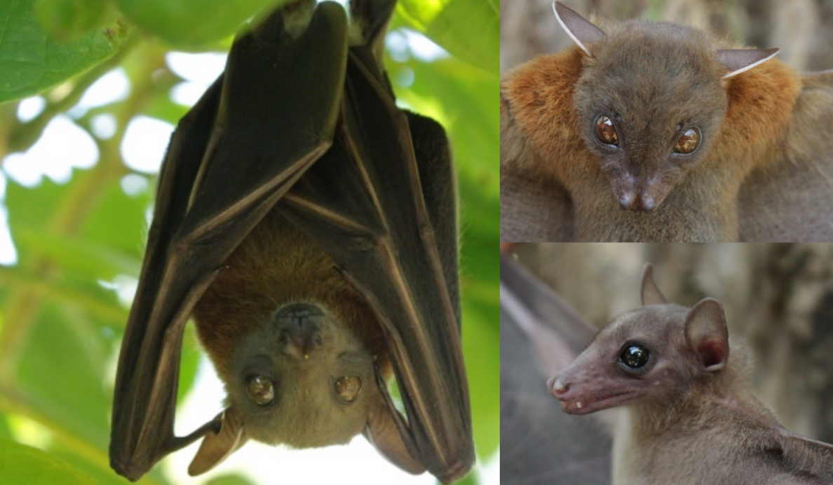
Initial patients with SARS had contact with game animals according to epidemiological studies. Animal traders had a higher prevalence of SARS-CoV IgG than the general population according to seroprevalence studies (Centers for Disease Control and Prevention, 2003). Himalayan palm civets and racoon dog in live animal markets were the first animals found to carry SARS-CoV-like viruses (Guan, 2003). Subsequent searches for the natural animal host in a surveillance study among non-caged animals from wild areas in Hong Kong revealed that a closely related bat coronavirus, SARS-related Rhinolophus bat coronavirus HKU3 (SARSr-Rh-BatCoV HKU3), existed in Chinese horseshoe bats (Rhinolophus sp.) (Lau, 2005). According to phylogenetic analysis, it is suggested that HCoV-NL63 may have originated from bat coronaviruses that have diverged 563-822 years ago (Huynh et al, 2012), while HCoV-HKU1 also likely originated from a bat coronavirus (Woo et al, 2009). The replicase gene of MERS-CoV, in its initial phylogenetic analysis showed that the virus was most closely related to bat CoV-HKU4 and CoV-HKU5 (Zaki et al, and Chan et al, van Boheemen et al, 2012). Phylogenetic analysis in recent studies using partial RdRp sequences showed that the MERS-CoV was more closely related to betacoronaviruses in bats from Europe and Africa (Cotten et al and Annan et al, 2013). Continued search for the natural definitive and intermediate animal hosts may help to halt the ongoing epidemic. After bats were identified as the natural host of SARS-CoV, the hunt for novel coronaviruses in bats accelerated . Bats are the only flying mammals capable of travelling long distances which facilitates transmission of viruses (Zhang et al and Chan et al, 2013). In addition to the SARSr-Rh-BatCoV HKU3, Bat coronaviruses identified in Hong Kong include the Rhinolophus bat CoV-HKU2 (Lau et al, 2007), Tylonycteris bat coronavirus CoV-HKU4 (Woo et al, 2006), Pipistrellus bat CoV-HKU5 (Woo et al, 2006), Myotis bat CoV-HKU6 (Woo et al, 2006), Miniopterus bat CoV-HKU7 (Woo et al, 2006) and Miniopterus bat CoV-HKU8 (Woo et al, 2006), Hipposideros and Rousettus bat CoV-HKU10 (Lau et al, 2012). Other bat coronaviruses have also been found in different parts of the world (Tong et al, 2009 and Yang et al, 2013). Birds are also an important origin and reservoir of emerging viruses causing human infections such as avian-origin influenza viruses other than bats (Chan et al, 2013). Numerous coronaviruses were also found in a wide range of birds (Woo et al, 2012). Bird coronaviruses identified in Hong Kong include bulbul CoV-HKU11, thrush CoV-HKU12, munia CoV-HKU13, white-eye CoV-HKU16, sparrow CoV-HKU17, magpie robin CoV-HKU18, night heron CoV-HKU19, wigeon CoV-HKU20, and common moorhen CoV-HKU21. Most of the bat coronaviruses belong to Alphacoronavirus and Betacoronavirus, while bird coronaviruses belong to Gammacoronavirus and Deltacoronavirus (Woo et al, 2010). Coronaviruses were also found in many other domestic and wild mammals, such as dogs, cats, pigs, whales and alpacas (Woo et al, 2012, Mihindukulasuriya et al, 2008, Crossley et al, 2012, and Perlman and Netland, 2009) besides bats and birds. Since the virus has originated from the Middle East, camels are suspected to be a reservoir of MERS-CoV and some patients had contacted with camels prior to symptom onset. Indeed, enteric coronaviruses have been identified in a camel previously (Wünschmann et al, 2002).
Risk Factors
MERS-CoV
Travel history to Arabian countries, animal-related exposure, food exposure, underlying health conditions and behaviors are the predictors for MERS-CoV. Significant factors include direct dromedary exposure in 2 weeks, concomitant with diabetes or heart disease, currently smoking tobacco (Alraddadi, 2016). Camel contact is a recognized risk factor for Middle East respiratory syndrome coronavirus (MERS-CoV) infection. Because specific camel exposures associated with MERS-CoV seropositivity are not fully understood, Ahmed et al (2019) investigated worker–camel interactions and MERS-CoV seroprevalence. In this study, on multivariable analyses, working as a camel salesman, handling live camels or their waste, and having diabetes were associated with seropositivity among all workers, whereas handling live camels and either administering medications or cleaning equipment was associated with seropositivity among market workers.
SARS-CoV
Severe acute respiratory syndrome (SARS) is caused by a novel coronavirus, transmitted from human to human by droplets or by direct contact. In general, people at greatest risk of SARS have had direct, close contact with someone who’s infected, such as family members and health care workers (MFMER, 2019). Airborne spread of the virus also accounted for certain community outbreaks of SARS (Yu et al. 2004). Accounting for one fifth of the global total, healthcare workers (HCW) were at the highest risk of having the disease (World Health Organization 2003). In Hong Kong, 23% of the cases of SARS were healthcare workers. In Canada and Singapore, the proportions were higher (40% and 41%, respectively), as they had fewer SARS cases in the community than Hong Kong. Risk factors for infection in HCWs have been studied extensively, and a review on SARS infection and healthcare workers disclosed a number of risk and protective factors (Chan-Yeung & Yu 2003; Lau et al. 2004). For example, lack of awareness and preparedness when the disease first struck, lack of training in infection control procedures, poor compliance with the use of personal protection equipment (PPE), poor institutional infection control measures, exposure to high-risk procedures such as intubation and nebulisation, and exposure to unsuspected SARS patients were associated with SARS infection. Measures to prevent nosocomial infection included establishing isolation wards for triage SARS patients; training and monitoring hospital staff in infection-control procedures; active and passive screening of HCWs; enforcement of droplet and contact precautions; and compliance with the use of PPE.
